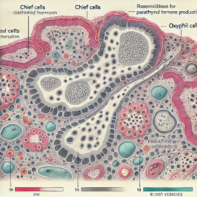
Here are some practical tips for identifying histology slides under a light microscope, focused specifically on understanding structural details. These techniques will help beginners and advanced users alike to recognize key features in different tissues.
1. Familiarize Yourself with Basic Tissue Types and Their Structure
- Epithelial Tissue: Look for closely packed cells with little to no space between them. Pay attention to the number of layers (single = simple; multiple = stratified) and cell shape (squamous, cuboidal, columnar).
- Connective Tissue: Identify loose or dense arrangements of cells with fibers (collagen, elastin) in between. Check for a matrix with cells spaced apart, often scattered with visible blood vessels.
- Muscle Tissue: Notice elongated cells. In skeletal muscle, cells are long and have striations (parallel bands). Cardiac muscle has striations with visible intercalated disks, and smooth muscle is smooth with no striations.
- Nervous Tissue: Recognize neurons by their large cell body and branching dendrites. Look for supporting glial cells, which appear smaller and more numerous around neurons.
2. Identify the Staining Patterns and Colors
- Hematoxylin and Eosin (H&E): This common stain colors nuclei blue (hematoxylin) and cytoplasm pink (eosin). The contrast helps in identifying cell boundaries and tissue architecture.
- Periodic Acid-Schiff (PAS): Highlights glycogen and carbohydrate-rich structures in magenta, useful for identifying mucus-secreting glands and basement membranes.
- Trichrome Stain: Stains muscle fibers red, collagen blue or green, and nuclei dark purple/black, helping distinguish between muscle, connective tissue, and other structures.
3. Examine the Cell Shape and Arrangement
- Cell Shape: Recognize the shape (e.g., round, elongated, polygonal) to identify tissue types. Squamous cells are flat, cuboidal are square, and columnar are tall.
- Cell Arrangement: Look for specific patterns:
- Glands: Round or oval clusters of cells indicate glandular tissue.
- Lining Tissues: Epithelial cells arranged in continuous sheets suggest lining of organs or surfaces.
- Clusters and Nodules: Often represent lymphoid tissue, like in lymph nodes or spleen.
4. Look for Specific Structural Markers
- Basement Membrane: A thin, dense layer beneath epithelial cells, best seen with PAS stain, providing support and barrier function.
- Lumen: A clear space surrounded by cells, seen in tubular structures like glands, blood vessels, and ducts.
- Intercellular Junctions: These may appear as darker, fine lines between cells, especially in epithelial and cardiac muscle tissues.
5. Identify Key Structural Features of Common Organs
- Liver: Look for hepatocytes arranged in rows with central veins. Identify sinusoids, the small spaces between hepatocyte rows.
- Kidney: Observe the renal cortex and medulla. Glomeruli (circular structures in the cortex) and tubules (elongated structures) are distinguishing features.
- Lung: Identify alveoli (small air sacs) with thin walls and occasional blood vessels or bronchioles.
- Intestine: Look for villi (finger-like projections) lined by epithelial cells with goblet cells (mucus-secreting cells) scattered within.
6. Observe the Fibrous and Extracellular Matrix Components
- Collagen Fibers: Thick, wavy fibers commonly found in connective tissues; they stain pink with H&E.
- Elastic Fibers: Thin, branched fibers, often found in tissues requiring flexibility (e.g., lungs, blood vessels), staining black or dark purple with special stains.
- Ground Substance: This often appears as a clear or light-staining background in loose connective tissue, giving the tissue a soft, gel-like structure.
7. Use Magnification Wisely
- Low Magnification (4x-10x): Start with low power to get a general view of the tissue structure and arrangement.
- High Magnification (40x-100x): Zoom in on specific areas to examine cellular details, nucleus shape, and cell arrangement more closely.
8. Practice Recognizing Artifacts
- Artifacts like folds, tears, or air bubbles can distort structures. Learn to identify and ignore these by focusing on consistent structural features that define the tissue.
9. Identify Surrounding Features for Context
- Often, structures like blood vessels, connective tissue, or nerve fibers are close to the main tissue type. These surrounding features can help confirm the tissue type (e.g., arteries and veins often accompany muscles).
Conclusion
Consistent practice and familiarization with these techniques will build strong foundational skills for accurate identification of histology slides.


0 Comments