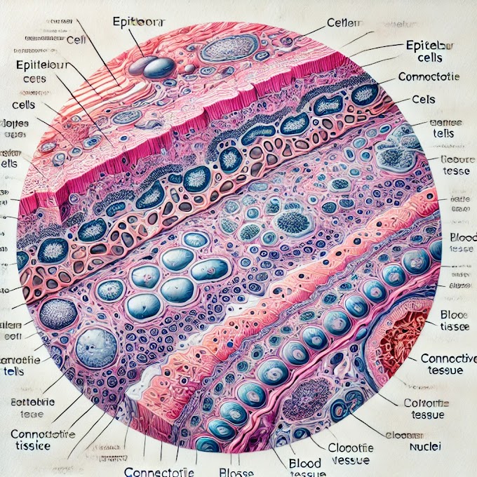
Histology Slide Identification Points
What is histology?
- Histology is the study of the microscopic structure of tissues, allowing us to understand the architecture and function of different cells and tissues in the body.
How can I identify different tissue types on a slide?
- Identifying tissue types involves recognizing certain characteristics such as cell shape, arrangement, staining patterns, and the presence of specific structures like glands or muscle fibers.
What are some common staining techniques used in histology?
- The most common stains include Hematoxylin and Eosin (H&E), Periodic acid–Schiff (PAS), and Trichrome. Each stain highlights specific cellular components and aids in tissue identification.
What is the purpose of H&E staining?
- H&E staining is used to distinguish between different tissue components: hematoxylin stains cell nuclei blue, while eosin stains the cytoplasm and extracellular matrix pink, helping differentiate between cell types and tissue structures.
What are the basic steps in identifying a histology slide?
- Start by examining the overall tissue architecture, identify any characteristic patterns, look at cell shapes and staining, and compare with known structures.
How can I distinguish between epithelial and connective tissues?
- Epithelial tissue has tightly packed cells with little extracellular matrix, often forming layers, whereas connective tissue has more extracellular matrix and supports or connects other tissues.
What are the key differences between skeletal, cardiac, and smooth muscle under a microscope?
- Skeletal muscle has striations and multiple peripheral nuclei, cardiac muscle has branching fibers and intercalated discs, and smooth muscle has spindle-shaped cells with central nuclei and no striations.
What should I look for when identifying nervous tissue?
- Look for neurons with prominent cell bodies and axons, and supportive glial cells. The organization of neurons and surrounding structures can also help differentiate CNS and PNS tissues.
Why are staining artifacts sometimes seen on histology slides?
- Artifacts can occur during slide preparation and may include folds, bubbles, or uneven staining. These should be distinguished from actual tissue features.
How can I improve my histology slide identification skills?
- Practice by examining a wide range of slides, comparing with reference images, and studying specific identification points for each tissue type.
Where can I find high-quality histology slide images for study?
- There are several online resources and atlases available, including digital slide libraries. You may also offer resources or images on your own website.
What tools are helpful for examining histology slides?
- A high-quality light microscope with appropriate magnification levels, digital slide viewers, and reference guides or atlases are essential tools.
Can I view histology slides online?
- Yes, many institutions and websites provide virtual histology slides that can be viewed under different magnifications, which are useful for remote learning.
How does slide preparation affect the appearance of tissues?
- Preparation steps like fixation, embedding, and staining can influence tissue appearance and introduce variations. Proper technique minimizes artifacts and improves clarity.
What is the best way to learn histology for exams or practicals?
- Combine theory with hands-on slide review, practice with identification quizzes, and use resources like atlases and digital slides to reinforce your understanding.


%20Volume.webp)


0 Comments