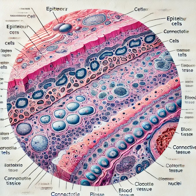
A comprehensive article outline on the microscopic study in anatomy, focusing on histology slides. This will cover all aspects, from defining microscopic features to explaining slide preparation, staining, and its significance for medical students.
Microscopic Study in Anatomy: A Guide to Histology Slides
Introduction
- Microscopy is essential in anatomical studies, particularly histology, which examines tissue structures on a microscopic level.
- Understanding microscopic anatomy helps medical students and professionals gain insights into tissue functions, cell interactions, and disease pathology.
1. What Are Microscopic Features in Histology?
- Definition: Microscopic features refer to the structures, cells, and subcellular components visible only under a microscope.
- Purpose: Observing these features allows a deep examination of tissues to understand their physiological and pathological states.
- Types of Microscopic Features:
- Cell Structure: Identification of cell types, nuclei, cytoplasm, and organelles.
- Tissue Architecture: The organization of cells in a tissue, essential for distinguishing between normal and abnormal tissue patterns.
- Intercellular Components: Fibers, ground substance, and other extracellular components critical for structural and functional integrity.
2. Why Is Microscopy Important in Medical Studies?
- Medical Diagnosis: Histological examination helps in diagnosing diseases, understanding tissue changes, and identifying abnormal growths.
- Educational Insight: For medical students, it’s essential for learning anatomy, pathology, and physiology, forming a bridge between structure and function.
- Research and Advancements: Understanding cellular and tissue morphology helps in developing treatments, drugs, and surgical techniques.
3. Overview of Slide Preparation: From Tissue Collection to Observation
- Preparing slides for microscopic study involves several steps to preserve and visualize the tissue effectively.
4. Tissue Collection and Fixation
Tissue Collection
- Sampling: Tissue samples can be obtained from biopsies, autopsies, or experimental procedures.
- Handling: Proper handling is essential to avoid distortion or damage to the tissue structure.
Fixation
- Purpose of Fixation: Fixation stabilizes and preserves tissue structure by halting decomposition and preventing cellular changes.
- Common Fixatives:
- Formalin: The most widely used fixative, providing excellent preservation of cell structure and protein.
- Glutaraldehyde: Used for electron microscopy due to its ability to crosslink proteins.
- Fixation Process:
- Immersion Fixation: Small tissue pieces are submerged in a fixative solution.
- Perfusion Fixation: Injecting fixative into the blood vessels, used primarily in research settings.
- Duration: Generally takes from several hours to overnight, depending on the tissue type and size.
5. Dehydration and Embedding: Preparing for Sectioning
Dehydration
- Purpose: Removing water from tissue is essential to allow it to be embedded in a wax-like medium.
- Process:
- Tissues are passed through graded alcohol solutions (e.g., 70%, 80%, 90%, and 100%) to gradually remove water.
- Often followed by a clearing agent like xylene to make the tissue transparent.
Embedding
- Embedding Medium: Commonly, paraffin wax is used; for electron microscopy, resin is preferred.
- Embedding Process: The tissue is placed in molten paraffin wax, then cooled to form a solid block that supports the tissue for slicing.
6. Sectioning: Creating Thin Slices for Microscopy
- Purpose of Sectioning: Sectioning allows tissue to be cut into thin slices (5–10 micrometers thick) for light microscopy.
- Microtome Usage: A microtome is used to slice the paraffin-embedded tissue into thin sections.
- Mounting on Slides: Sections are placed on glass slides with adhesives to ensure they remain stable during staining and observation.
7. Staining: Enhancing Contrast for Observation
- Purpose of Staining: Cells and tissues are transparent, so stains are applied to increase contrast, making structures more visible.
- Types of Stains:
- Hematoxylin and Eosin (H&E): The most common stain, where hematoxylin stains nuclei blue and eosin stains cytoplasm pink.
- Special Stains: Periodic Acid-Schiff (PAS) for carbohydrates, Masson’s Trichrome for connective tissue, etc.
- Staining Process:
- Deparaffinization: Removal of paraffin by soaking the slides in xylene.
- Rehydration: Passing slides through graded alcohol solutions back to water for staining.
- Staining Steps: Each dye is applied in a specific order, sometimes followed by counterstaining to enhance contrast.
- Mounting and Coverslipping: A coverslip is added over the stained tissue with a mounting medium for preservation and observation.
8. Observing and Analyzing Slides Under the Microscope
- Light Microscope Usage: Adjust magnification, focus, and light intensity to study structures at various levels.
- Analyzing Different Tissues:
- Epithelial Tissues: Look for layers and shapes of cells.
- Connective Tissues: Identify fiber types and matrix components.
- Muscle Tissues: Distinguish between striated and smooth muscle based on organization.
- Nervous Tissue: Recognize neurons and supporting cells.
9. Preservation and Maintenance of Prepared Slides
- Storage: Slides should be kept in dust-free slide boxes or cabinets to protect from contamination.
- Labeling: Proper labeling with sample information, date, and stain type aids in future reference.
- Preventing Fading: Certain stains may fade over time; controlled storage conditions, such as low light and temperature, help extend longevity.
10. Why Histology is Important for Medical Students
- Foundational Knowledge: Histology links cellular structure to function, providing essential insights into how the body works.
- Pathology Understanding: A strong foundation in histology allows medical students to recognize pathological changes in tissues, critical for diagnosing diseases.
- Skill Development: Learning to prepare, stain, and analyze tissue slides enhances students' practical and observational skills.
- Application in Clinical Practice: Understanding tissue structure and function is essential for interpreting biopsies, surgeries, and laboratory findings.
Conclusion
- The microscopic study of anatomy through histology slides is fundamental to medical education, research, and practice.
- From tissue fixation and sectioning to staining and observation, each step reveals unique insights into the human body, laying the groundwork for understanding health, disease, and medical interventions.


0 Comments