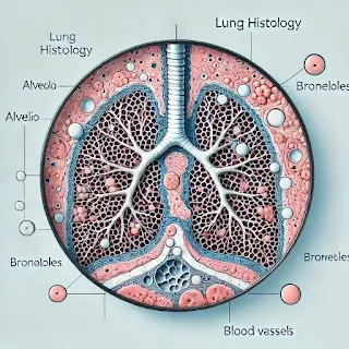
A lung histology slide contains the following labeled areas with distinct markings and colors:
Labeling and Markings:
Red Arrows:
- Labeled as "Alveolar sac & ducts":
- Points to alveolar structures, which are key sites for gas exchange.
- Labeled as "Bronchiole":
- Indicates the bronchiole, an airway leading to the respiratory bronchioles and alveoli.
- Key Description:
- The bronchiole is a tubular structure lined with simple columnar or cuboidal epithelium.
- Alveolar sacs and ducts appear as open spaces surrounded by thin septal walls.
- Labeled as "Alveolar sac & ducts":
Blue Arrow:
- Labeled as "Alveoli & simple squamous epithelium":
- Highlights the alveoli, which are lined by simple squamous epithelial cells (Type I pneumocytes).
- Key Description:
- Alveoli are thin-walled spaces for gas exchange.
- The simple squamous epithelium facilitates the diffusion of oxygen and carbon dioxide.
- Labeled as "Alveoli & simple squamous epithelium":
Purple Arrow:
- Labeled as "Proximal bronchiole":
- Refers to the proximal section of the bronchiole leading into the smaller respiratory bronchioles.
- Key Description:
- Proximal bronchioles have a thicker wall with smooth muscle and lack cartilage or glands.
- Labeled as "Proximal bronchiole":
Orange Arrow:
- Labeled as "Lymphatic nodules":
- Points to lymphatic tissue within the lung section.
- Key Description:
- Lymphatic nodules are part of the immune defense, located in the interstitial spaces or near airways.
- Labeled as "Lymphatic nodules":
Summary:
Each colored arrow corresponds to a specific lung histology feature critical for understanding lung anatomy and function:
- Red arrows: Bronchioles and alveolar ducts/sacs.
- Blue arrow: Alveoli and simple squamous epithelium.
- Purple arrow: Proximal bronchiole.
- Orange arrow: Lymphatic nodules.
Under a light microscope, a lung histology slide typically reveals the following key structures and identification points:
1. Alveoli:
- Description: Small, thin-walled sacs forming the main site for gas exchange.
- Appearance: Empty, honeycomb-like spaces surrounded by thin septa.
- Key Features: The alveolar walls contain capillaries and elastic fibers. The lining includes:
- Type I alveolar cells: Thin, squamous epithelial cells aiding gas exchange.
- Type II alveolar cells: Cuboidal cells producing surfactant, found at the corners of alveoli.
2. Alveolar Septa:
- Description: The walls separating adjacent alveoli.
- Appearance: Thin layers containing capillaries, fibroblasts, and elastic fibers.
- Key Features: Often show nuclei of endothelial cells and interstitial tissue.
3. Bronchioles:
- Description: Small airways leading to alveolar ducts.
- Appearance: Tubular structures lined with cuboidal or columnar epithelium.
- Key Features:
- Lack cartilage and glands.
- Smooth muscle layer visible surrounding the bronchiolar lumen.
4. Blood Vessels:
- Description: Pulmonary capillaries and arteries associated with gas exchange.
- Appearance: Capillaries are seen as thin, small channels interwoven in the alveolar walls.
- Key Features:
- Pulmonary arteries appear larger with thicker walls.
- Veins are found within the connective tissue.
5. Respiratory Bronchioles and Alveolar Ducts:
- Description: Transitional structures between bronchioles and alveoli.
- Appearance: Respiratory bronchioles have some alveoli opening directly into their walls.
- Key Features:
- Alveolar ducts are elongated passages with alveoli along their walls.
6. Pleura:
- Description: The outermost lining of the lung.
- Appearance: A thin, fibrous connective tissue layer with a mesothelial covering.
- Key Features: Visible as a smooth layer at the periphery of the lung section.
7. Interstitial Tissue:
- Description: Connective tissue supporting the lung structure.
- Appearance: Contains fibroblasts, collagen, and elastic fibers.
- Key Features: May show lymphatic vessels and occasional immune cells.
Each of these structures plays a crucial role in lung function, and the marked region in your histology slide may correspond to one or more of these features depending on the area of interest.
Overview of the lungs, covering their anatomy, physiology, biology, histopathological features, and clinical significance:
1. Anatomy of the Lungs
The lungs are paired respiratory organs located in the thoracic cavity. They are vital for oxygen exchange in the body.
Gross Anatomy:
- Lobes: The right lung has three lobes (superior, middle, and inferior), while the left lung has two (superior and inferior) to accommodate the heart.
- Trachea and Bronchi:
- The trachea bifurcates into the right and left bronchi, which further divide into smaller bronchioles and alveolar ducts.
- Pleura:
- A double-layered membrane:
- Visceral pleura (covers the lungs).
- Parietal pleura (lines the thoracic cavity).
- A double-layered membrane:
- Blood Supply:
- Pulmonary arteries carry deoxygenated blood to the lungs for oxygenation.
- Pulmonary veins return oxygenated blood to the heart.
- Lymphatic System:
- Lymph nodes are located around the hilum for immune defense and drainage.
Microscopic Anatomy:
- Alveoli: Tiny air sacs where gas exchange occurs.
- Bronchioles: Small airways lacking cartilage, lined with cuboidal epithelium.
- Respiratory Epithelium:
- Ciliated columnar cells, goblet cells (mucus production), and basal cells.
2. Physiology of the Lungs
The lungs are essential for maintaining gas exchange and acid-base balance.
- Gas Exchange:
- Oxygen from inhaled air diffuses into alveolar capillaries, binding to hemoglobin.
- Carbon Dioxide from blood diffuses into alveoli to be exhaled.
- Ventilation Mechanics:
- Inhalation: Diaphragm contracts, increasing thoracic volume and reducing pressure to draw in air.
- Exhalation: Diaphragm relaxes, allowing elastic recoil to expel air.
- Regulation of pH:
- By controlling CO2 levels, the lungs help maintain blood pH through the bicarbonate buffer system.
- Immune Function:
- The lungs trap and remove foreign particles via mucus and ciliary action.
3. Biological Features
- Cell Types:
- Type I Alveolar Cells: Thin squamous cells for gas exchange.
- Type II Alveolar Cells: Cuboidal cells producing surfactant to reduce surface tension.
- Macrophages: Engulf pathogens and debris within the alveoli.
- Surfactant:
- A lipid-protein mixture essential for preventing alveolar collapse.
- Immune Cells:
- Mast cells and lymphocytes provide defense against inhaled pathogens.
4. Histopathological Features
Diseases of the lungs often show specific structural changes:
- Normal Histology:
- Thin alveolar walls, clear alveolar spaces, and healthy capillary networks.
- Pathological Conditions:
- Pneumonia:
- Inflammation of alveoli filled with pus or fluid.
- Histology: Alveolar exudates with neutrophils and bacteria.
- Emphysema:
- Destruction of alveolar walls, leading to enlarged airspaces.
- Histology: Loss of alveolar septa and reduced capillary networks.
- Fibrosis:
- Thickening of alveolar walls due to collagen deposition.
- Histology: Dense fibrotic tissue and inflammatory infiltrates.
- Lung Cancer:
- Abnormal proliferation of epithelial cells (e.g., adenocarcinoma or squamous cell carcinoma).
- Histology: Disorganized growth with nuclear atypia and increased mitotic activity.
- Pneumonia:
5. Clinical Significance
The lungs are susceptible to various diseases due to their constant exposure to environmental agents.
Common Diseases:
- Asthma:
- Chronic airway inflammation leading to wheezing and dyspnea.
- Pathophysiology: Hyperresponsiveness of bronchial smooth muscle.
- Chronic Obstructive Pulmonary Disease (COPD):
- Includes emphysema and chronic bronchitis.
- Leads to reduced airflow and oxygen exchange.
- Lung Cancer:
- Often linked to smoking and environmental pollutants.
- Common types: Small cell lung cancer and non-small cell lung cancer.
- Pneumonia:
- Infection causing alveolar inflammation and fluid accumulation.
- Pulmonary Fibrosis:
- Scarring of lung tissue, leading to stiffened lungs and reduced compliance.
- Asthma:
Diagnostic Tools:
- X-rays: Identify abnormalities like fluid, masses, or infections.
- CT Scans: Provide detailed imaging for tumors and interstitial diseases.
- Pulmonary Function Tests (PFTs): Measure airflow and lung capacity.
- Bronchoscopy: Allows direct visualization of airways.
Treatment Approaches:
- Oxygen therapy for hypoxia.
- Medications: Bronchodilators, corticosteroids, and antibiotics.
- Surgical options: Lobectomy or lung transplantation for severe cases.


0 Comments