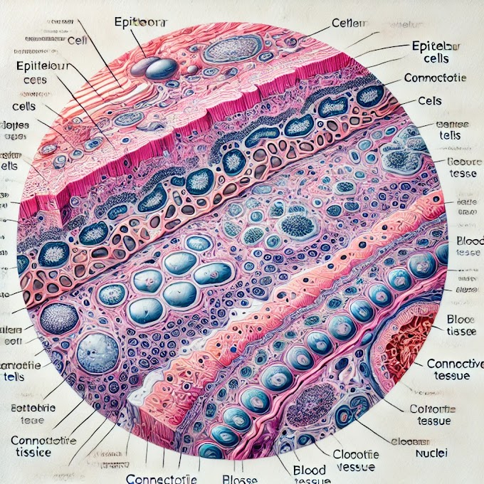
In a histology slide of lip tissue, several key layers and structures are typically visible:
1. Epithelium (Stratified Squamous Epithelium)
- Outer Layer: This is the surface layer that provides a protective barrier. In the lip, it’s often keratinized or partially keratinized in the outer region (vermilion border) to withstand friction.
- Basal Layer: The deepest part of the epithelium, where new cells are generated and gradually pushed up toward the surface.
2. Dermis
- Connective Tissue: Just below the epithelium, this layer contains fibrous connective tissue, supporting and connecting the different components. Collagen and elastin fibers are prevalent here.
- Blood Vessels: The dermis has a rich blood supply that helps nourish the tissue, including smaller blood capillaries near the epithelium, which contribute to the lip's red color.
- Nerve Endings: Responsible for sensory input, such as touch and pain, and giving the lips their sensitivity.
3. Muscle Layer (Orbicularis Oris Muscle)
- This layer consists of skeletal muscle tissue, mainly the orbicularis oris muscle, which is responsible for lip movement. Muscle fibers appear as elongated, striated cells aligned in bundles.
4. Submucosa (If Present)
- In some parts of the lip, a submucosal layer with minor salivary glands might be seen. These glands contribute to the moisture of the lip tissue.
Each of these layers plays a distinct role, working together to provide structure, flexibility, sensitivity, and moisture to the lips.
Here’s an overview of the lip’s anatomy, physiology, histopathology, and clinical significance:
Anatomy
The lips are soft, mobile structures that form the anterior part of the mouth. Each lip has three main regions:
- External Skin: Composed of keratinized epithelium, like typical skin.
- Vermilion Border: The reddish, transitional zone between the outer skin and inner mucous membrane.
- Inner Mucous Membrane: A non-keratinized mucosal layer, continuous with the lining of the mouth.
Key structures include:
- Epithelium: Stratified squamous epithelium with both keratinized and non-keratinized regions.
- Connective Tissue: Includes blood vessels, nerves, and connective fibers.
- Muscles: The orbicularis oris muscle, which allows for movements like speaking, smiling, and kissing.
- Minor Salivary Glands: Found in the inner lining and contribute to moisture.
Physiology
The lips have several important functions:
- Sensory Perception: Rich in nerve endings, lips are extremely sensitive, allowing for tactile sensation, temperature detection, and pain perception.
- Articulation and Expression: Lips are essential for speech and non-verbal communication. The orbicularis oris muscle enables precise movements for facial expressions and articulation of sounds.
- Protective Barrier: The lips act as a barrier to pathogens and regulate moisture, with sebaceous glands contributing to natural lubrication.
Histopathology
Histopathology of the lips involves examining tissue changes under a microscope, which can reveal various conditions:
- Inflammatory Conditions: Such as cheilitis (inflammation of the lips) or angular cheilitis, which affects the corners of the mouth.
- Precancerous and Cancerous Lesions: Actinic cheilitis (often due to UV exposure) is a precancerous condition. Squamous cell carcinoma can also arise from chronic sun damage or tobacco use.
- Infectious Conditions: Viral infections like herpes simplex virus (HSV) present with blistering on histology slides, showing multinucleated cells and characteristic viral inclusions.
- Autoimmune Conditions: Conditions like lupus erythematosus can cause characteristic changes in lip tissue.
Clinical Significance
Common Lip Disorders:
- Herpes Labialis: Caused by HSV, leading to painful blisters.
- Angular Cheilitis: Inflammation at the lip corners, often due to fungal or bacterial infections or nutritional deficiencies.
- Actinic Cheilitis: Chronic sun exposure can lead to this condition, which may progress to squamous cell carcinoma.
- Lip Cancer: Often linked to UV exposure, tobacco, and alcohol use, particularly affecting the lower lip.
Diagnostics:
- Biopsy: In suspicious lesions, a biopsy helps confirm diagnosis, especially for malignancies or precancerous lesions.
- Histopathology: Essential in identifying structural changes due to disease or trauma.
Treatment Approaches:
- Surgical Excision: Common for lip cancers and persistent lesions.
- Topical or Systemic Therapies: For conditions like herpes or fungal infections, topical antivirals or antifungals may be used.
- Photoprotection: Sun protection measures, like lip balms with SPF, are recommended to prevent actinic cheilitis and reduce cancer risk.
Summary
The lips, while simple in appearance, play vital roles in communication, sensation, and protection. Awareness of their anatomy and pathologies is crucial in detecting diseases early, especially for cancerous and infectious conditions that may initially appear benign.


0 Comments