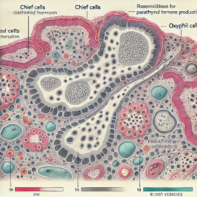
histology slide of the ureter, the following key structures and layers are typically identified:
1. Mucosa Layer
- Transitional Epithelium: The innermost lining of the ureter is composed of transitional epithelium, which is unique because it can stretch and accommodate the movement of urine. The transitional epithelium appears in multiple layers:
- Basal Layer: Contains small, cuboidal cells close to the basement membrane.
- Intermediate Layer: Has larger, polygonal cells above the basal layer.
- Superficial (Apical) Layer: Contains large, rounded cells called "umbrella cells," which help protect underlying cells from the urine's osmotic effects.
2. Lamina Propria
- Directly beneath the transitional epithelium is the lamina propria, a connective tissue layer providing support and nutrition to the epithelium. It contains blood vessels, nerves, and loose connective tissue.
3. Muscularis Layer
- The muscularis layer surrounds the mucosa and is composed of smooth muscle fibers arranged in two main layers:
- Inner Longitudinal Muscle Layer: Smooth muscle fibers running along the length of the ureter.
- Outer Circular Muscle Layer: Smooth muscle fibers arranged in a circular manner around the ureter.
- Together, these muscle layers help propel urine from the kidneys to the bladder through coordinated contractions (peristalsis).
4. Adventitia
The outermost layer of the ureter, composed of loose connective tissue. It anchors the ureter to surrounding structures and contains blood vessels, nerves, and lymphatics. In certain areas closer to the bladder, the adventitia may be replaced by a serosa layer (a thin epithelial covering) if it is within the peritoneal cavity.Anatomy of the Ureter
The ureter is a muscular tube approximately 25-30 cm in length, extending from the renal pelvis to the bladder. It has three primary layers:
- Mucosa: Lined with transitional epithelium, allowing it to stretch as urine flows.
- Muscularis: Composed of smooth muscle with an inner longitudinal and an outer circular layer. This enables peristaltic movement.
- Adventitia: The outermost layer of connective tissue that anchors the ureter to surrounding structures.
The ureters enter the bladder obliquely, which creates a valve-like mechanism preventing backflow, protecting the kidneys from high bladder pressures.
Physiology of the Ureter
The ureter’s primary physiological function is to propel urine from the kidneys to the bladder. This process is achieved through coordinated peristaltic waves in the muscularis layer, regulated by autonomic nerves. Each wave moves urine downward and prevents reflux, ensuring unidirectional flow.
The ureter’s transitional epithelium and its protective umbrella cells form a barrier against urine’s high osmolarity, shielding the tissue from chemical injury.
Histopathology of the Ureter
The histopathological examination of the ureter involves studying changes in its cellular structure under various pathological conditions:
- Ureteritis: Inflammation of the ureter, often due to infection or injury, can lead to infiltration by inflammatory cells, thickening of the mucosa, and edema.
- Ureteral Fibrosis: Chronic inflammation can cause fibrosis or scarring of the ureteral wall, potentially obstructing urine flow.
- Urothelial Carcinoma: Cancerous growths may arise from the transitional epithelium. Microscopically, these appear as abnormal cell proliferation with a loss of normal cellular organization.
- Ureteral Strictures: Narrowing of the ureter due to injury, inflammation, or congenital abnormalities may be identified through thickening or fibrosis on histology slides.
Clinical Significance of the Ureter
Ureteral Obstruction: Commonly caused by kidney stones, tumors, or strictures, an obstruction prevents urine flow from the kidney to the bladder. This can lead to hydronephrosis (swelling of the kidney) and kidney damage if left untreated. Treatment options range from ureteral stents to surgical intervention.
Ureteral Calculi (Stones): Stones can form in the ureter and block urine flow, causing severe, colicky pain often referred to as “ureteral colic.” Imaging and sometimes histopathology can confirm the presence and type of stones, which may require surgical removal or lithotripsy.
Congenital Abnormalities: Conditions like duplication of the ureter or ectopic ureter (where the ureter doesn’t insert into the bladder properly) can lead to recurrent infections, obstruction, or incontinence, often detected in childhood.
Ureteral Cancer: Urothelial carcinoma of the ureter is relatively rare, but early detection is critical for effective treatment. Patients may present with hematuria (blood in urine), back pain, or frequent urinary infections.
Reflux Nephropathy: In cases where the valve mechanism between the bladder and ureter fails, urine may flow backward into the kidneys. This condition, known as vesicoureteral reflux, can increase the risk of kidney infections and scarring, particularly in children.
Summary
The ureter’s anatomy and physiology enable it to function as a robust urine conduit, while its unique histological features provide protection and adaptability. Its clinical relevance is underscored by its involvement in conditions such as obstruction, stones, cancer, and congenital anomalies. Understanding ureteral structure, function, and pathology is crucial in diagnosing and managing urinary tract disorders effectively.
written by: Ikrambaigtech
Youtubchannel subscribe


0 Comments