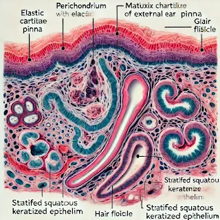
Under The Light Microscopic Structure
1. Stratified Squamous Keratinized Epithelium (Yellow arrow):
·
This outer layer covers the
external ear. It consists of multiple layers of cells that help in protecting
the underlying tissue from damage, dehydration, and pathogens.
2.
Hair Follicle (Orange arrow):
·
Hair follicles are embedded
in the skin of the external ear. They house the root of the hair and play a
role in skin health and sensory functions.
3. Gland (Red
arrow):
·
These glands are likely
sebaceous glands, responsible for secreting oil (sebum) to lubricate and
protect the skin of the pinna.
4. Perichondrium (Green arrow):
·
This is the dense
connective tissue that surrounds the elastic cartilage. It provides nutrients
to the cartilage and plays a vital role in cartilage growth and repair.
5. Matrix, Elastic Fibers, and Lacuna (Purple arrow):
·
The elastic cartilage
within the external ear is composed of a matrix rich in elastic fibers, giving
it flexibility. Lacunae are small spaces where cartilage cells (chondrocytes)
are located.
6. Connective Tissue and Gland (Blue arrows):
·
The underlying connective
tissue houses glands and supports the structure of the pinna, contributing to
the ear's flexibility and durability.
This slide highlights the structural components that allow the pinna to maintain its shape and perform its functions in sound collection and protection.
For an OSPE or practical exam, here are 4 key identification points for the external ear pinna histology slide:
- Stratified Squamous Keratinized Epithelium
- Perichondrium
- Elastic Fibers in the Cartilage Matrix
- Hair Follicle
These points can help students focus on identifying major features without needing additional explanations
Here is the labeled histology slide of the external ear pinna, showcasing key structures like the perichondrium,
elastic fibers, stratified squamous keratinized epithelium, hair follicles,
glands, and more.
Complete
Guide for Medical Students:
Anatomy of the External Ear (Pinna):
- External
ear primarily consists of elastic cartilage covered by skin.
The cartilage provides structural support and flexibility, enabling the
pinna to retain its shape.
- Perichondrium:
A dense layer of connective tissue surrounding the cartilage, essential
for the nourishment of the cartilage.
- Elastic
cartilage: Contains elastic fibers in a collagen matrix that gives
flexibility and resilience to the ear.
- Stratified
Squamous Keratinized Epithelium: The outermost layer of skin covering
the pinna, providing protection from external elements.
- Hair
follicles and glands (sebaceous and sweat glands): Found in the skin
layer, these structures maintain skin health and function.
Physiology:
The external ear is designed to capture sound waves and
direct them into the ear canal. The elasticity of the pinna allows it to modify
its shape and adjust to different frequencies of sound. The cartilage prevents
damage by absorbing minor shocks.
Histopathology:
- Normal
elastic cartilage shows a regular arrangement of elastic fibers within
the matrix. Abnormalities in these fibers can indicate diseases like chondritis
(inflammation of cartilage).
- Histological
stains reveal the presence of elastic fibers, which appear dark under
specific staining techniques.
Clinical Significance:
- Chondritis:
Infection or inflammation of the ear cartilage, often resulting from
trauma or ear piercings.
- Perichondritis:
Involves inflammation of the perichondrium, leading to painful swelling.
- Cauliflower
ear: Repeated trauma to the ear may cause a collection of blood
between the cartilage and the perichondrium, leading to permanent
deformity if untreated.
This comprehensive guide can help your MBBS students
understand both the structure and function of the external ear, as well as its
clinical relevance
Click Here to Watch Youtube Video


0 Comments