

Under The Light Microscopic View
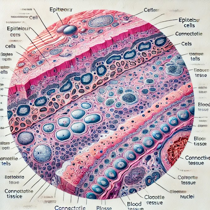
This histology slide of transitional epithelium with labeled key features. Here’s a description of each marked area:
Multi-cell Layer:
- Basal Layer: The innermost layer (closest to the basement membrane), consisting of cuboidal or columnar cells. These cells are generally small and help anchor the epithelium to the underlying tissue.
- Middle Layer: This layer consists of pear-shaped or rounded cells, which provide structural support and allow for flexibility.
- Surface Layer: The outermost layer, with dome-shaped or umbrella cells. These surface cells can flatten and stretch, which is important for accommodating changes in volume, especially in organs like the bladder.
Star-shaped Lumen:
- The lumen is the cavity or space inside the tubular structure where fluids pass. The star-shaped appearance is characteristic of the relaxed state of transitional epithelium, often seen in organs like the ureter or bladder.
Basement Membrane:
- The basement membrane is a thin layer of extracellular matrix that anchors the epithelial cells to the underlying connective tissue. It provides structural support and acts as a barrier while allowing selective permeability of substances between the epithelium and connective tissue.
Overview of Transitional Epithelium
- Location: Transitional epithelium lines the urinary system organs, such as the bladder, ureters, and part of the urethra.
- Function: Its primary function is to allow these organs to expand and contract as they fill and empty. The unique structure of its cells allows it to stretch without losing integrity or creating gaps.
- Special Features: This epithelium can transition between a relaxed state (thicker with dome-shaped surface cells) and a stretched state (thinner with flattened surface cells).
Transitional epithelium is critical for maintaining a barrier in regions of the urinary system that undergo frequent distension, providing a balance of protection and flexibility.
1. Cellularity:
Epithelial tissues are densely packed with cells, forming continuous sheets.
2. Polarity:
Cells have distinct apical (top) and basal (bottom) surfaces, each serving specific functions.
3. Attachment:
Epithelial cells adhere to a basement membrane, a structural foundation supporting the tissue.
4. Arrangement:
Cells can be single-layered (simple) or multi-layered (stratified), influencing their protective capabilities.
5. Shapes:
Cells may exhibit squamous (flat), cuboidal (cube-shaped), or columnar (elongated) morphology, reflecting their specialized roles.
6. Specializations:
Microvilli, cilia, or goblet cells enhance functions like absorption, movement, or secretion.
7. Layers:
Simple epithelia have one cell layer, while stratified epithelia have multiple layers, affecting durability.
Understanding these features aids in precise identification when examining histology slides.
overview of transitional epithelium, including its anatomy, physiology, biochemistry, histopathology, and clinical significance.
1. Anatomy
- Location: Transitional epithelium, also known as urothelium, lines organs of the urinary system that experience significant stretching, such as the urinary bladder, ureters, and part of the urethra.
- Cell Layers: It consists of multiple cell layers that vary in appearance depending on the degree of stretch.
- Basal Layer: Contains cuboidal or columnar cells that are attached to the basement membrane. This layer is responsible for cell regeneration and anchors the epithelium to the underlying connective tissue.
- Intermediate Layer: Contains pear-shaped or rounded cells. These cells act as transitional support between the basal and superficial layers.
- Surface Layer: Composed of large, dome-shaped cells, also known as “umbrella cells,” which can stretch and flatten as needed.
- Star-shaped Lumen: When relaxed, the lumen (inner cavity) lined by transitional epithelium often appears star-shaped due to the folds in the epithelium.
2. Physiology
- Stretching Ability: Transitional epithelium can expand and contract without tearing or creating gaps between cells. This allows organs like the bladder to hold varying amounts of urine.
- Barrier Function: It acts as a protective barrier, preventing pathogens, toxins, and urine from passing into underlying tissues.
- Surface Protection: The cells of the surface layer have specialized proteins, such as uroplakins, that form plaques to protect the cells from the toxic effects of urine.
3. Biochemistry
- Uroplakins: These are specialized proteins found in the apical (top) layer of umbrella cells. They form a tough, impermeable surface, which protects the epithelium from the acidic and potentially toxic components of urine.
- Cell Junctions: Transitional epithelial cells are tightly connected by cell junctions, particularly tight junctions and desmosomes, which maintain the integrity of the tissue and prevent urine from leaking into underlying tissues.
- Cell Regeneration: Basal cells continually divide to replace cells shed from the surface layer, ensuring tissue integrity and protection.
4. Histopathology
- Normal Histology: In a healthy state, transitional epithelium shows a well-defined structure with a basal layer, intermediate layer, and dome-shaped surface cells. The tissue appears multi-layered and can be thicker when relaxed or thinner when stretched.
- Pathological Changes:
- Cystitis: Inflammation of the bladder, often caused by a bacterial infection, which leads to damage of the transitional epithelium. Chronic cystitis can cause hyperplasia (increased cell proliferation) and sometimes squamous metaplasia (change of cell type).
- Transitional Cell Carcinoma (TCC): This is the most common cancer of the urinary tract. It typically arises from the urothelium of the bladder but can also affect the ureters and renal pelvis. TCC may present as papillary or flat growths and can invade underlying tissues if not treated early.
- Squamous Metaplasia: Chronic irritation, often from repeated infections or bladder stones, can lead to a transformation of the transitional epithelium into a squamous (flattened) epithelium. While this may be a protective response, it can also increase the risk of malignancy.
- Leukoplakia: A condition where thickened, white patches form on the transitional epithelium, typically due to chronic irritation. This is sometimes seen in smokers or patients with chronic bladder inflammation.
- Urothelial Dysplasia: Abnormal growth and appearance of cells in the transitional epithelium, often a precursor to carcinoma in situ or invasive cancer.
5. Clinical Significance
- Urinary Tract Infections (UTIs): UTIs are common infections that can affect the transitional epithelium lining of the bladder (cystitis) or ureters (ureteritis). Recurrent UTIs can lead to chronic inflammation and may predispose patients to epithelial changes or cancer.
- Bladder Cancer: Transitional cell carcinoma (TCC) of the bladder is the most common type of bladder cancer. Risk factors include smoking, exposure to certain chemicals (like those used in the dye and rubber industries), and chronic bladder irritation.
- Bladder Dysfunction: Conditions like interstitial cystitis and overactive bladder can cause chronic pain and urge to urinate frequently. Changes in the transitional epithelium can sometimes be found in these conditions, although the exact mechanisms are not fully understood.
- Protective Adaptations: The ability of transitional epithelium to adapt to varying volumes in the urinary tract is crucial for preventing urine leakage into the bloodstream and surrounding tissues. This adaptability and impermeable barrier are important for maintaining urinary system health.
- Urine Cytology: Transitional epithelial cells are often examined in urine cytology, a test used to detect cancer cells or abnormalities in the urinary tract. Urine samples are analyzed for exfoliated cells from the transitional epithelium to identify signs of cancer or infection.
Summary
Transitional epithelium, found in the urinary system, is a specialized, multi-layered epithelium that allows stretching and contraction. It plays an essential role in protecting underlying tissues from the potentially harmful components of urine and is highly adaptable. Histologically, it consists of basal, intermediate, and surface (umbrella) cells. Pathologically, changes in the transitional epithelium can lead to diseases like transitional cell carcinoma, cystitis, and squamous metaplasia. Understanding this epithelium is critical for diagnosing and managing urinary tract diseases, particularly bladder cancer and chronic infections.
Click here to watch videos on YouTube channel ikrambaig@tech
Written by: ikrambaigtech
YouTube video
Download video


LinkedIn page 📄📃👇 image


0 Comments