

Under The Light Microscopic View
%20bone%20histology%20slide,%20showing%20labeled%20structures%20including%20periosteum,%20bony%20trabeculae,%20osteon,%20p.webp)
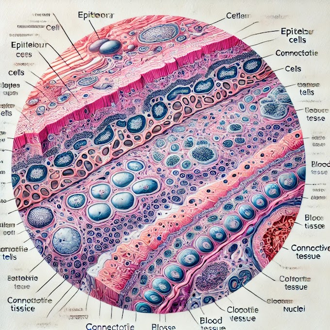
you can identify various structures and features that contribute to its overall composition. Here are some key points to consider:
Periosteum:
The external surface of the bone is covered by periosteum.Periosteum comprises of an external stringy layer and an internal cell layer. It may not be however particular in that frame of mind as it seems to be in reduced bone.Endosteum:
The inward surface of the trabeculae is fixed with endosteum, a cell layer that adds to bone renovating.
Bone Marrow:
Springy bone contains red bone marrow, answerable for hematopoiesis (platelet arrangement). Search for regions with an organization of hematopoietic cells, adipocytes (fat cells), and veins.Compact Bone (possibly in some areas):
Compact bone might be available in specific areas, particularly at the external edges or regarding the trabeculae. Smaller bone has a more coordinated structure with osteons and lamellae.
Osteon (Haversian System):
Unlike compact bone, spongy bone does not have well-defined osteons. Spongy bone has a cross section like construction called trabeculae, which gives strength and backing.Osteoblasts and Osteoclasts:
Recognize osteoblasts, which are liable for bone development. Search for osteoclasts, which are engaged with bone resorption.Primitive (Undifferentiated) Cells:
A few regions could contain crude or undifferentiated cells that can lead to different bone cell types.
Read more about spongy bone,
Identifying histological features on a spongy bone (also called trabecular or cancellous bone) slide involves examining the tissue under a microscope.
Trabeculae:
- Thin, branching bony plates that make up the spongy or cancellous bone.
- Form a lattice-like structure that provides support and helps resist stress.
Bone Marrow Spaces:
- Spaces between trabeculae filled with bone marrow.
- Red bone marrow is responsible for hematopoiesis (blood cell formation), while yellow bone marrow stores adipocytes (fat cells).
Red Marrow Spaces:
- Regions of the spongy bone containing red bone marrow.
- More prevalent in certain bones and responsible for blood cell formation.
Osteocytes:
- Mature bone cells embedded in lacunae within the trabeculae.
- Connected to each other and to the central canal (canaliculi) by processes in the bone matrix.
Lacunae:
- Small cavities or spaces in the bone matrix where osteocytes are located.
Canaliculi:
- Microscopic channels that connect adjacent lacunae and allow communication between osteocytes.
- Facilitate the exchange of nutrients and waste products.
Endosteum:
- A thin layer of connective tissue that lines the surfaces of trabeculae and the medullary cavity.
- Contains osteoprogenitor cells involved in bone remodeling and repair.
Periosteum:
- The outer covering of bones, but it may not be visible in spongy bone sections.
- Composed of connective tissue that provides support and contributes to bone repair.
Blood Vessels:
- Identify blood vessels within the bone marrow spaces, supplying nutrients and oxygen to bone cells.
Trabecular Thickness and Arrangement:
- Note the thickness and arrangement of trabeculae, which can vary between bones and locations within bones.
Recollect that the particular attributes noticed may differ relying upon the area of the bone (long bone, level bone, and so forth) and the age of the person from whom the bone example was taken. Moreover, histological staining procedures can improve the perceivability of specific designs.
Overview of spongy bone
1. Anatomy of Spongy Bone
Structure:
- Also known as cancellous or trabecular bone, spongy bone is a porous, less dense type of bone found primarily in the ends of long bones (epiphyses), within the interior of vertebrae, and inside flat bones like the ribs, pelvis, and skull.
- Unlike compact bone, which has a solid, continuous structure, spongy bone consists of a lattice-like network of trabeculae (small, bony rods or plates).
- These trabeculae are organized in a way that provides strength while keeping the bone lightweight, with spaces between them filled with red or yellow bone marrow.
Locations:
- Epiphyses of long bones
- Inside vertebral bodies
- Ribs, pelvis, sternum, and skull bones
2. Physiology of Spongy Bone
Functions:
- Support and Protection: Spongy bone helps absorb shock and reduce stress on the bone, especially during movement or weight-bearing activities.
- Bone Marrow Storage: The spaces between trabeculae house bone marrow, where hematopoiesis (production of blood cells) occurs.
- Metabolic Role: The trabeculae allow for a high surface area, facilitating mineral exchange, especially for calcium and phosphate.
- Adaptability: Spongy bone can remodel itself quickly in response to stress, optimizing bone density and structure based on activity levels.
3. Biology and Composition of Spongy Bone
Cell Types:
- Osteoblasts: Cells that build new bone matrix by producing collagen and aiding in mineralization.
- Osteoclasts: Cells that break down bone tissue, balancing bone formation and resorption.
- Osteocytes: Mature bone cells embedded in the bone matrix, maintaining bone tissue and detecting mechanical stress.
Matrix Composition:
- Organic Components: Primarily collagen fibers that provide flexibility and tensile strength.
- Inorganic Components: Mostly calcium and phosphate minerals in the form of hydroxyapatite, which give the bone its hardness and rigidity.
Blood Supply:
- Blood vessels penetrate through the bone marrow spaces within the trabeculae, providing essential nutrients and oxygen to the bone cells.
4. Histology of Spongy Bone
Microscopic Structure:
- Trabeculae: Thin plates of bone forming a meshwork. They do not contain osteons (the structural unit of compact bone) but have lamellae (layers) similar to those in compact bone.
- Bone Marrow: The spaces between trabeculae are filled with either red marrow (involved in hematopoiesis) or yellow marrow (primarily composed of fat).
- Endosteum Lining: The trabeculae are lined by a thin layer of connective tissue called the endosteum, which contains bone-forming cells like osteoblasts and osteoclasts.
Bone Cells:
- Trabeculae are lined with osteoblasts on the surface and contain embedded osteocytes, while osteoclasts are often found on the surface, where bone resorption occurs.
5. Clinical Significance of Spongy Bone
Bone Disorders and Conditions:
- Osteoporosis: In osteoporosis, bone density decreases, affecting spongy bone more severely than compact bone. This makes bones, especially in the spine and hips, more susceptible to fractures.
- Bone Marrow Disorders: Since spongy bone contains red marrow, conditions affecting blood cell production, such as leukemia or anemia, are associated with this area.
- Bone Cancer: Certain cancers, such as multiple myeloma and bone metastases, often involve spongy bone due to its high vascularity.
- Osteomyelitis: Spongy bone can be susceptible to infections due to its vascularity and proximity to bone marrow.
- Fractures: The lattice structure of spongy bone helps absorb impact, but severe trauma can still lead to fractures. Spongy bone typically heals faster than compact bone due to its rich blood supply.
Therapeutic Applications:
- Bone Marrow Transplant: Bone marrow extracted from the spongy bone of donors is used in bone marrow transplants to treat blood disorders.
- Calcium Homeostasis: The spongy bone serves as a primary site for the storage and release of minerals, which is crucial for maintaining blood calcium levels.
6. Histopathology of Spongy Bone
Histopathological examination of spongy bone can reveal signs of various diseases:
- Osteoporosis: Thin, less dense trabeculae, with reduced bone volume and weakened structure.
- Osteopetrosis: Excessively dense trabeculae due to the failure of osteoclasts to resorb bone properly.
- Paget’s Disease: Abnormal trabecular patterns, increased osteoblastic and osteoclastic activity, and mosaic bone formation.
- Bone Tumors: Abnormal cell proliferation, invasion into trabeculae, and possible formation of atypical cells.
Diagnostic Tools:
- Bone Biopsy: A sample of spongy bone may be taken to assess marrow conditions.
- Imaging: X-rays, CT scans, and MRIs provide detailed visualization of the trabecular structure and can show areas affected by disease.
In summary, spongy bone plays a critical role in both structural support and physiological functions such as mineral homeostasis and blood cell production. Its histopathological study aids in diagnosing various bone-related diseases and disorders, making it a significant area in both anatomy and clinical medicine.
written by: Ikrambaigtech.blogspot.com
you can watch videos slides on my youtube channel ikrambaig@tech
YouTube channel video

LinkedIn page click below 👇 image

📌 Pinterest page 📄📃 below 👇 image


Free video download click here


0 Comments